Computed tomography, or CT, is a ubiquitous X-ray imaging technique used to perform more than 300 million medical imaging exams globally each year. Use of the technique continues to grow, with CT increasingly employed as a first-line diagnostic tool for conditions such as coronary artery disease, as well as moving into preventive care and early detection, such as lung cancer screening.
But there are still limitations to what conventional CT can achieve in the clinic. For starters, it’s not always possible to image all patients: medical implants, for example, can create image artefacts that inhibit diagnostic accuracy. There are also shortfalls with regard to image reproducibility and standardization, both essential for ongoing evaluation of disease progress. Finally, there’s a growing need for functional data alongside the standard anatomical CT images.
“These three shortcomings are the ones we had in mind when we started moving into a new era of how CT imaging can be provided,” said Philipp Fischer, head of CT at Siemens Healthineers. “This was the starting point for our thoughts around Naeotom Alpha.”
The culmination of more than 15 years of research, Naeotom Alpha is the world’s first photon-counting CT scanner. Photon-counting technology enables dramatic improvements in diagnostic imaging, including increased resolution and a reduction in radiation dose by up to 45% over conventional CT detectors. Naeotom Alpha is now cleared for clinical use in the USA and Europe, and Siemens Healthineers will be showcasing its new system at next week’s RSNA Annual Meeting.
Crystal breakthrough
Speaking at a media briefing, Fischer described the main differences between conventional and photon-counting CT technology. Conventional CT requires two conversion steps to turn photons into a medical image: X-rays are collected by a scintillator, which uses many photons together to generate an optical signal; a photodiode then converts this light into an electrical signal. The process is stable and reliable; but two-step conversion limits the dose efficiency and, with many photons contributing to the optical signal, the achievable resolution is limited.
“With photon-counting technology, we move from a two-step conversion process to a single one-step conversion of X-ray photons into an electrical current that generates the medical image,” Fischer explained, noting that removing a conversion step increases dose efficiency. “The large difference is that with this new technology, we are able to assess each and any single photon separately, and also assess the energy level of each and any photon.”
This new approach to image acquisition, however, required the development of an entirely new detector material: high-purity cadmium telluride (CdTe) crystals. CdTe crystals deliver the highest spatial resolution of any CT imaging system to date, said Fischer, enabling pixels nine times smaller than used in conventional CT, without any dose penalty.
The crystals also completely eliminate electronic noise from the detector, resulting in a higher signal-to-noise ratio and lower dose requirements. Another advantage is that, unlike conventional CT that often underrepresents low-energy photons, photon-counting technology uses all photons equally, which should be particularly beneficial for soft-tissue imaging. Finally, the detector’s intrinsic spectral sensitivity means that spectral data are available in every scan.
Clinical gains
Fischer shared some examples of how these CT advances could prove invaluable in the clinic. When diagnosing coronary disease, for instance, seven million invasive catheterization procedures are performed each year, half of which do not lead to therapy. CT angiography provides a non-invasive diagnostic alternative. But conventional CT scans suffer from image artefacts due to calcifications or implanted stents or pacemakers, precluding a large patient population from this approach.
The spectral imaging offered by photon-counting technology enables differentiation between calcifications, stents, vessel walls and contrast media. This allows the removal of unwanted data from the image, increasing diagnostic accuracy. “With photon-counting technology, we have the goal to make non-invasive coronary imaging available to all patients that could benefit from it,” said Fischer.
In oncology, meanwhile, Naeotom Alpha’s high precision is particularly beneficial. Cancer patients undergo diagnostic scanning and many follow-up exams to assess treatment response and disease progression. With conventional CT, however, the image acquisition parameters can influence the image itself, preventing accurate comparison of scans over time. Photon-counting CT, on the other hand, provides consistent signal quality, with stable Hounsfield unit values for each patient across every scan.
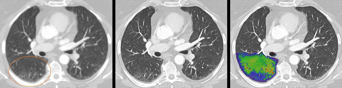
Another target application is lung imaging, where multiple images are often required to achieve a meaningful diagnosis. “With conventional CT, you need to know in advance what you are looking for, and then design your imaging exam,” Fischer explained. “You cannot look for all the detail in one exam without compromising on patient dose, resolution or functional image information.” But with photon-counting technology, a single scan can provide both structural images and functional information on the perfusion of the lung.
Initial experience
The dual-source Naeotom Alpha scanner is already installed at 22 sites. One of the first installations was at the University Hospital Augsburg in Germany, where the system has been in routine clinical use since April 2021.
To date, the Naeotom Alpha has been used to scan more than 4000 patients, in areas including oncology, neurology, cardiology and musculoskeletal imaging. “We are very happy with the results so far,” said Thomas Kröncke, director of the hospital’s department of diagnostic and interventional radiology.
“The important thing to me is seeing better and seeing more, due to the intrinsic spectral separation that photon counting allows,” said Kröncke. “Spectral post-processing is always possible, and the considerable increase in spatial resolution and higher intrinsic iodine contrast make it possible to reduce not only the radiation but also the contrast material.”