- Science News
- Frontiers news
- Research integrity: A closer look at gel and Western Blot image cropping
Research integrity: A closer look at gel and Western Blot image cropping
Author: Bushra Khair, Research Integrity Specialist
Since last year, the Frontiers Research Integrity team has followed up on more than 4,000 manuscripts containing cropped gel and Western Blot images which required additional scrutiny.
In this blog, we take a closer look at gel and Western Blot image cropping. Figures and images are a key component of many research papers. It is important, then, that authors submit figures correctly and to the standards expected of quality scientific literature. Oftentimes, that is the case. But from time to time, we do encounter issues**.**
What are Gels and Western Blots?
Gels and Western Blots are used in molecular biology laboratories to identify and analyze macromolecules such as nucleic acids and proteins in a sample. Gels are often used for studying nucleic acids while Western Blots are used to visualize and study proteins. For example, if researchers investigating pancreatic cancer suspect a particular protein might be involved in the disease, a series of Western Blot experiments could help to compare the expression of the protein in both cancer cells and normal healthy cells. The experiments might detect the protein being expressed in cancer cells, but not healthy cells and vice versa, helping researchers to understand the disease further. Both of these experiments involve preparing the samples in question, followed by gel electrophoresis. For Western Blot experiments, the separated proteins are then transferred onto a nitrocellulose or PVDF membrane for further preparation with antibodies specific to the proteins of interest and finally analyzed in a special imaging machine.
Once complete, a digital image is then captured and saved to present later on, often in a manuscript. Below is an example of a full length, unedited original Western Blot image:
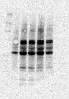
Figure 1: Unedited Western Blot image
It is common practice to crop a gel or Western Blot image to present in research publications. The cropping process removes irrelevant parts of the captured image and presents only the proteins of interest. In some cases, the membrane itself may be spliced after protein transfer and staining and then probed with antibodies to reduce the volume of reagents needed for the experiment. However, a gel itself is never spliced. Only the captured image may be cropped to only present the area of interest.
Here is an example of acceptable Western Blot image splicing, where irrelevant parts of the Western Blot images were cropped and presented to make the splicing clear to readers:

Figure 2: Acceptable Western Blot image splicing
Here is an example of acceptable Western Blot cropping, where the membrane itself has been spliced (original and unedited):

Figure 3: Acceptable Western Blot membrane cropping
Problematic cropping cases
Gel and Western Blot cropping can be problematic. In cases where duplicated features are detected or inappropriate editing is applied, red flags are raised about the integrity of the image. This can occur when parts of the image, such as specific bands or lanes are cropped, extracted, and then pasted onto another area of the image. This is often referred to as splicing. Such cases are of concern when cropping the captured gel or Western Blot image changes the scientific inferences made from the image. It is best practice to save un-edited, originally captured image files so they are available to share upon request.
In the Frontiers Research Integrity department, we ensure that published images maintain strong scientific integrity. This is achieved in collaboration with our assigned Editors who provide insight, using their specific expertise to help guarantee the scientific integrity and quality of Frontiers journals. As a member of the Committee on Publication Ethics, we also apply the appropriate guidelines and policies to ensure that cropped gel and Western Blot images do not contain manipulated features, which ultimately, would change the conclusion of the article.
When conducting quality checks on manuscripts containing cropped gel and Western Blot images, we ask authors to provide their raw and originally saved image files for evaluation. The commonly accepted file type is TIF, however, TIFF and JPEG are also accepted. More information can be found in our author guidelines.
It is important that authors label these image files correctly, according to the nucleic acid or protein of interest and that these are aligned with the images presented in the manuscript. It is also important to declare any membrane splicing to clarify cases where a full-length Western Blot image is not provided. In cases where raw, unedited images cannot be provided, the manuscript will be withdrawn as this data is not considered to be reproducible. However, authors are always welcome to resubmit once these experiments are repeated or if the original image files are retrieved. While it is common practice to include cropped Western Blot images in manuscripts so as to include the relevant proteins, cases where there is questionable cropping, undeclared cropping or duplicate bands raise concerns for the team. In such cases, the authors will be requested to provide originally saved image files for assessment.
Here is an example of unacceptable gel and Western Blot cropping, where features have been spliced and manipulated. The image on the right is a marked version with colored squares presenting duplicated bands that have been cropped out and pasted onto the blot:
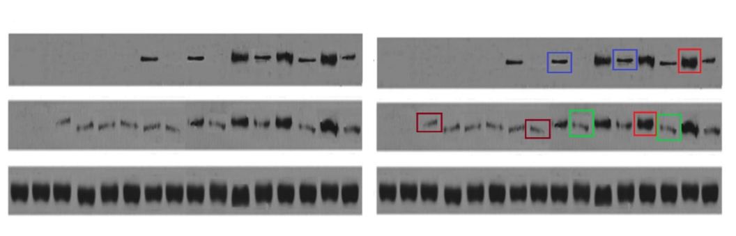
Figure 4: Unacceptable Western Blot cropping
At Frontiers, the Research Integrity team also uses our Artificial Intelligence Review Assistant (AIRA) to screen potentially fraudulent images. AIRA detects and flags duplicate images within figures of manuscripts. The Research Integrity team then assesses these flagged images and use their expertise to decide whether the case requires further attention and clarification from the authors.
Throughout academic publishing, gel and Western Blot cropping is a common practice, and in many cases, horizontal and vertical cropping is acceptable. However, authors must take care and ensure that any cropping is declared in figure legends. It is also crucial that authors check that any cropping or edits applied do not damage the scientific integrity of the data presented in these images. As mentioned, it is always good practice to keep raw image files stored securely in case these are requested by the Research Integrity office or by future readers. Maintaining good practices directly contributes to the quality of scientific publications, journals and reflects positively on scientists. It also helps to establish trust in the paper’s conclusions because the research will be regarded as reliable, and may then be used to help inform or build further studies.
_Bushra Khair is currently an Editor Outreach Coordinator, having joined Frontiers in 2019 as a Research Integrity Specialist. She graduated from Queen Mary University London with an M.Sc in Cancer and Molecular Pathology and Genomics in earlier that year. Bushra also holds a 1st class B.Sc in Human Nutrition from the University of Westminster where she graduated in 2018.
Her interests lie in molecular biology and is drawn towards learning more specifically about molecular pathology and how these processes lead to cancers. Since joining Research Integrity, Bushra has become increasingly interested in tackling image manipulation and falsification. Overall, she is driven to ensure that those involved in scholarly publishing adhere to ethical practices. In her current role as an Editor Outreach Coordinator, Bushra is committed to ensuring that Editors who join Frontiers are high-quality researchers who adhere to ethical publication practices._