
Proton MR spectroscopy can identify changes in the brains of people with multiple sclerosis (MS), according to a study published in Radiology. The findings could translate to earlier diagnosis and treatment, senior author Wolfgang Bogner of the Medical University of Vienna says in a statement released by the university.
“If confirmed in longitudinal clinical studies, this new neuroimaging technique could become a standard imaging tool for initial diagnosis, for disease progression and therapy monitoring of multiple sclerosis patients, and, in concert with established MRI, might contribute to neurologists’ treatment strategies,” he says.
MS affects the central nervous system and can cause fatigue, pain and coordination problems, the group notes. Almost 3 million people around the world suffer from the condition, and there’s no cure – only treatments such as physical therapy and medication to slow its progression.
One of the ways MS manifests is as white-matter lesions in the brain, which are associated with a loss of myelin, a protective coating around white matter fibres. But these lesions represent macroscopic damage, notes lead author Eva Heckova and colleagues: it would benefit patients to be able to identify earlier, microscopic changes in the brain suggestive of MS.
That’s why proton MR spectroscopy shows promise, since it can identify substances produced by a person’s metabolism that could indicate the presence of MS.
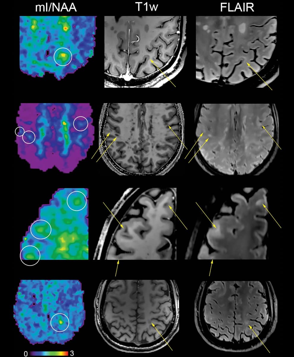
In the current study, proton MR spectroscopy was performed on a 7-tesla scanner using a technique called free-induction decay MR spectroscopic imaging, with 2 × 2 mm in-plane spatial resolution. For the study, the investigators used the technique to compare differences in normal-appearing white matter and cortical grey matter in the brains of 65 MS patients and 20 healthy controls.
The group found the following brain differences in patients with MS:
- Lower levels of an amino acid derivative called N-acetylaspartate, which is associated with compromised integrity of neurons.
- Higher levels of myo-inositol, which is involved in cell signalling; increased levels indicate significant inflammatory disease activity.
- Metabolic changes in normal-appearing white matter and cortical grey matter.
The brain changes this MR technique illuminated could have important clinical implications, according to Heckova.
“Some neurochemical changes, particularly those associated with neuroinflammation, occur early in the course of the disease and may not only be correlated with disability, but also be predictive of further progression such as the formation of multiple sclerosis lesions,” she says.
In an accompanying commentary, Peter Barker of Johns Hopkins University cautioned that although proton MR spectroscopy could be an effective way to identify MS earlier, it may be difficult to incorporate it into clinical practice.
“The number of 7-tesla scanners worldwide is tiny compared with 1.5 or 3-tesla, and the cost of installing and operating these systems is steep,” he says. “Thus, for the near future at least … both MRI and MR spectroscopy at 7-tesla are likely to be mainly of use for research studies in MS, where the best possible resolution and highest information content are needed.”
- This article was originally published on AuntMinnieEurope.com ©2022 by AuntMinnieEurope.com. Any copying, republication or redistribution of AuntMinnieEurope.com content is expressly prohibited without the prior written consent of AuntMinnieEurope.com.