RSNA 2021, the annual meeting of the Radiological Society of North America, brings together radiologists, radiation oncologists, medical physicists and related scientists to discuss the latest radiology research and developments. This year’s meeting is being held as a hybrid event, with more than 19,000 attendees expected to attend in-person in Chicago and another 4000 joining virtually. Here’s a small selection of the studies being presented at the conference this week, focusing on the latest innovative brain imaging research.
Mild to moderate COVID-19 during pregnancy doesn’t harm baby’s brain
Pregnant women appear to be more vulnerable to the SARS-CoV-2 virus, but little is known about the potential impact on an unborn child if the mother contracts COVID-19 during pregnancy. A study from Ludwig Maximilian University of Munich concludes that mild to moderate COVID-19 in pregnant women does not affect foetal brain development.
“Women infected with SARS-CoV-2 during pregnancy are concerned that the virus may affect the development of their unborn child, as is the case with some other viral infections,” explains senior author Sophia Stöcklein. “So far, although there are a few reports of vertical transmission to the foetus, the exact risk and impact remain largely unclear. The aim of our study was to fill this gap in knowledge regarding the impact of a maternal SARS-CoV-2 infection on foetal brain development.”
The researchers used foetal MRI to study 33 pregnant patients with COVID-19, acquiring T2-, T1- and diffusion-weighted brain images. The women were between 18 and 39 weeks into their pregnancies, with symptom onset at between four to 34 weeks.
Two expert radiologists evaluated the scans and quantitatively assessed structures of the brain stem and posterior fossa. They concluded that brain development – including brain surface and folding, cerebellar size and the size of all brain stem structures – was age-appropriate in all cases. There were no calcifications, signs of swelling or widening of the brain’s fluid-filled spaces.
Stöcklein cautions that the study only included mothers with mild to moderate symptoms and without hospitalization, emphasizing the importance of active protection against COVID-19 during pregnancy. The team plan to follow-up all newborns over the next five years, performing detailed neonatal assessment, and assessing neurological development.
Brain complications could affect over one in 100 hospitalized COVID-19 patients
A multi-institutional study has found that roughly one in 100 patients hospitalized with COVID-19 are likely to develop complications of the central nervous system. The retrospective study analysed nearly 40,000 hospitalized COVID-19 patients from seven US and four European university hospitals.
“Much has been written about the overall pulmonary problems related to COVID-19, but we do not often talk about the other organs that can be affected,” says lead author Scott Faro from Thomas Jefferson University. “Our study shows that central nervous system complications represent a significant cause of morbidity and mortality in this devastating pandemic.”
The patients in the study had an average age of 66 years old and many had comorbidities such as hypertension, cardiac disease and diabetes. The most common cause of hospital admission was confusion and altered mental status, followed by fever. Just over 10% of the cohort underwent neuroimaging; from these patients’ MRI or CT brain scans, the researchers identified 442 acute neuroimaging findings that were likely associated with COVID-19.
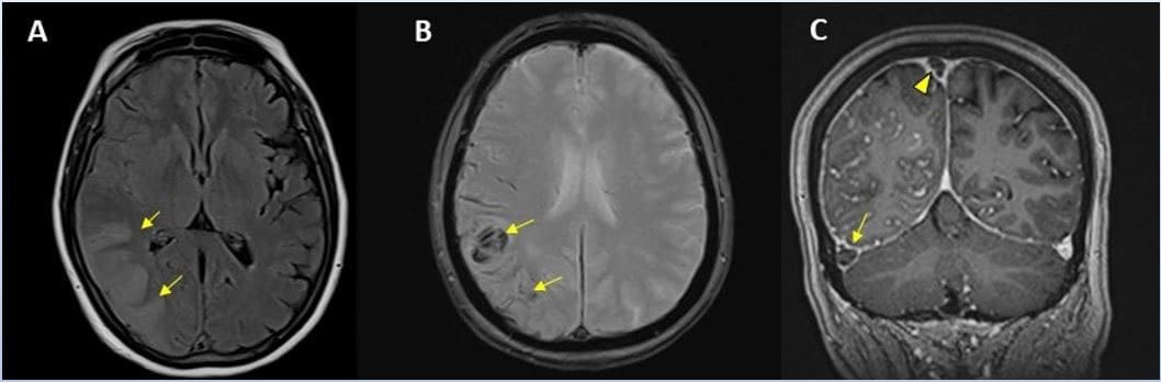
The overall incidence of central nervous system complications in all patients was 1.2%, suggesting that just over one in 100 patients admitted to hospital with COVID-19 will likely have a brain complication of some sort. The most common complication seen was ischemic stroke, followed by intracranial haemorrhage and then encephalitis (brain inflammation).
“It is important to know an accurate incidence of all the major central nervous system complications,” says Faro. “There should probably be a low threshold to order brain imaging for patients with COVID-19.”
MRI reveals how alcohol exposure impacts the foetal brain
Consuming alcohol during pregnancy can lead to foetal alcohol spectrum disorders, a range of conditions that may result in physical, behavioural and learning problems. In the first MRI-based study of pre-natal alcohol exposure, researchers have used MRI to identify early changes in the brain structure of foetuses exposed to alcohol.

“There are many post-natal studies on infants exposed to alcohol,” says Gregor Kasprian from the Medical University of Vienna. “We wanted to see how early it’s possible to find changes in the foetal brain as a result of alcohol exposure.”
Kasprian and colleagues studied 500 pregnant women referred for foetal MRI. Anonymous questionnaires revealed that 51 of the women had consumed alcohol during their pregnancy. The team analysed a final group of 24 foetuses with pre-natal alcohol exposure and a control group of 52 gender- and age-matched foetuses without alcohol exposure. At the time of imaging, the gestational age was between 20 and 37 weeks.
The researchers employed advanced post-processing techniques to generate super-resolution brain MR images and performed semi-automated atlas-based segmentation of the resulting images.

Pre-natal alcohol exposure alters functional connectivity
Analysis of 12 different foetal brain structures revealed increased volumes in the corpus collosum, the main connection between the brain’s two hemispheres, and decreased volumes in the periventricular zone, compared with controls. They note that this is the first time that a pre-natal imaging study has quantified these early alcohol-associated changes.
“It appears that alcohol exposure during pregnancy puts the brain on a path of development that diverges from a normal trajectory,” says Kasprian. “Foetal MRI is a very powerful tool to characterize brain development not only in genetic conditions, but also acquired conditions that result from exposure to toxic agents.”