Researchers at University of Illinois Urbana-Champaign have developed a novel trimodal brain imaging system, incorporating electroencephalography (EEG), functional magnetic resonance imaging (fMRI) and a third technique, event-related optical signal (EROS) imaging, reporting their findings in Human Brain Mapping.
The development of multimodal neuroimaging
Brain function was first measured almost 100 years ago using crude electrodes placed on the human scalp. Since then, EEG has become a routine clinical and research tool, penetrating the electrophysiology of the human brain. EEG measures the change in potential at the scalp, caused by the firing of neurons within the cortex, with millisecond temporal resolution. However, this signal results from a smeared summation of signals from within the brain, due to the different conductivities of the scalp, skull and cerebral-spinal fluid. This means that EEG cannot precisely trace signals back to the region of the brain that produced them.
By 1990, functional neuroimaging reached high spatial resolution with the development of fMRI, allowing brain activity to be traced to its origin with millimetre precision. fMRI measures the blood oxygenation level-dependent (BOLD) response, which relates the change in oxygenation of cortical blood to brain activity. As this method probes the haemodynamic response of the brain, rather than the electrophysiology, there is an inherent time delay of approximately 5–8 s between activation and the measured BOLD signal.
More recently, fMRI-compatible EEG instrumentation has facilitated simultaneous EEG and fMRI, capitalizing on the temporal resolution of EEG and the spatial resolution of fMRI. However, how the signals from these two modalities relate to one another is poorly understood.
Introducing EROS
EROS uses near-infrared light sources and detectors placed on the scalp to measure changes in ion transport during neural activity. Ion transport causes neurons to swell and shrink, subsequently scattering the light. By measuring these changes in light scattering, the technique can infer the location and temporal characteristics of cortical activity. While stand-alone EROS suffers from low sensitivity, incorporating it with EEG and fMRI clarifies the links between these two different measures.
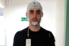
To create a proof-of-concept trimodal system, the scientists integrated EROS sensors (based on optical fibres and photomultiplier tube light detectors) into an MRI-compatible EEG–EROS cap. The cap, seen in the image above, does not produce any artefacts during the scanning.
The team tested this novel system using an emotional oddball experiment. This required participants to identify “oddball” circles presented on a screen in a string of standard (scrambled) and distracter (emotional and neutral) images. This type of stimulus is known to induce different spatial and temporal responses, making it ideal for testing the trimodal system. The researchers note that EROS is expected to be spatially similar to the fMRI data and correspond temporally with EEG.
Simultaneous EEG and fMRI measure sleep ‘inertia’
The EROS results identified a similar spatial response to the emotional and target stimuli in the frontal cortex as the fMRI, but provided a temporal resolution similar to EEG, whose signals originated from a different location in the brain. These findings demonstrate that introducing a third simultaneous imaging modality can expand our understanding beyond that which bimodal investigations are able to achieve.
The researchers highlight the considerable advantages of the trimodal system: the ability to overcome the limitations of each imaging modality; clarifying the links between each measure; and providing an excellent platform for future investigations into the spatial and temporal characteristics of brain mechanisms associated with healthy function and disease.