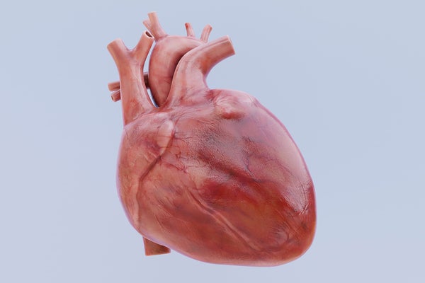
From artificial heart valves to cellular transplants, new treatments for cardiovascular ailments are being developed every day. To model how they work, researchers need a reliable way to observe the heart in action. Animal studies, computer models and various laboratory simulators made with dead heart tissue can all provide different views, but these approaches can be expensive, lacking in complexity or limited in their shelf life.
So to tinker with the heart, scientists have now developed a beating, biorobotic replica that can simulate the workings of both a healthy organ and a diseased one. This simulator combines pig heart tissue and soft robotic muscles and was described in two recent studies.
“Imagine a beating heart on a lab bench,” says Ellen Roche, a biomedical engineer at the Massachusetts Institute of Technology and the studies’ senior author. The simulator, which pumps a clear fluid instead of blood, is hooked up to instruments that measure blood flow, blood pressure, and more. It’s also customizable: the user can change the heart rate, blood pressure and other parameters, then watch how these changes affect the heart’s function in real time through an internal camera.
On supporting science journalism
If you're enjoying this article, consider supporting our award-winning journalism by subscribing. By purchasing a subscription you are helping to ensure the future of impactful stories about the discoveries and ideas shaping our world today.
The simulator accurately replicates how blood flows through the heart—something that existing benchtop simulators using dead heart tissue could not do. Using live heart tissue from a pig instead, animated by robotics, granted Roche’s team far more control. (Pig hearts are similar in size and layout to human hearts and are often used in research.) The new hybrid simulator can also last longer than a live organ that is used on its own: whereas a pig heart hooked up to a pump in the lab would only continue beating for a few hours, Roche’s team was able to keep the simulator’s synthetic muscles going for months. The researchers haven’t measured the simulator’s exact limits yet. “We need to do robust shelf-life fatigue testing to see exactly how many cycles these things can do,” Roche says.
When it comes to modeling blood flow through the heart, the left and right sides of the organ are each their own challenge. “They require very customized models,” Roche says. The researchers tackled the left side first by focusing on the mitral valve, which controls the flow between the left atrium and ventricle (the heart’s upper and lower chambers). They re-created the healthy motion of this system before modeling a condition in which the valve becomes leaky, called mitral regurgitation. To demonstrate that the model could be used as an accurate simulation, the team had cardiac surgeons correct the valve with three different surgical interventions. These results were described in one of the two recent studies, which was published on Wednesday in Device.
“It’s a very complex pumping motion that you have to create in order for the blood to pump to your body at a really high pressure and flow,” says Clara Park, who co-authored both studies as a Ph.D. student at Roche’s lab at M.I.T.
Next the team modeled the mechanics on the right side of the heart. “The right heart is kind of the thinner, weaker muscle,” Park says, and it “doesn’t pump as hard.” The right heart simulator can replicate both healthy and abnormal functioning. These results were described in the team’s other recent study, which was published last month in Nature Cardiovascular Research.
Sarah Vigmostad, a biomedical engineer at the University of Iowa, who was not involved in the papers, believes that these simulators would be valuable for surgical planning, training or educational purposes. “I can also imagine their value in testing new interventions ... designed to treat mitral regurgitation or other valve diseases,” she says. The ability to “tune” the heart to replicate various diseases could be very useful in research as well, she adds.
The biorobotic approach relies on live animal tissue, but Roche dreams of a completely 3-D-printed synthetic heart. Such an organ could be far more customizable. It could even be used to create patient-specific models that would allow people in treatment to observe a replica of their own beating heart in action and that would guide their doctors’ decisions. “We are moving to fully synthetic models [and] multi-material prints,” she says, which will require replicating heart tissue itself in the lab. “There's a lot of ongoing work.”