This year, researchers have once again had to deal with conferences going virtual and bouts of working from home. But that hasn’t stopped the medical physics community from continuing to develop and investigate advanced healthcare techniques and tools. Alongside ongoing efforts to help detect, analyse and prevent the spread of the SARS-CoV-2 virus, 2021 has seen also the introduction of novel cancer treatments, advances in diagnostic imaging technology and innovative new biomedical devices. Here are just a few of the research highlights that caught my eye this year.
Antibody therapy delivered directly into the brain
For the first time ever, researchers have delivered antibody therapy directly into the brain, to target breast cancer metastases. A team at Sunnybrook Health Sciences Centre used MR-guided focused ultrasound to non-invasively and temporarily open the protective blood–brain barrier (BBB), enabling the monoclonal antibody trastuzumab to reach specific areas of the brain. The researchers report the results from the first four patients in an ongoing clinical trial, all of whom had breast cancer with brain metastases.
The team used Insightec’s ExAblate device to perform transcranial focused ultrasound while simultaneously delivering intravenous trastuzumab. The drug was radiolabelled with 111In, enabling it to be visualized using SPECT. After BBB opening, the SPECT images showed that trastuzumab precisely targeted the brain tumours. MR imaging revealed that patients’ tumours shrank by between 7% and 31%, at up to four months after treatment. Importantly, the procedure caused no serious adverse effects.
Clinical linac produces FLASH beam
A team from Dartmouth has developed a method to convert a standard clinical linear accelerator (linac) used for radiation therapy to deliver a FLASH-capable, ultrahigh-dose rate radiotherapy beam. The process, which uses existing accessories, takes only 20 minutes to perform, or to reverse.
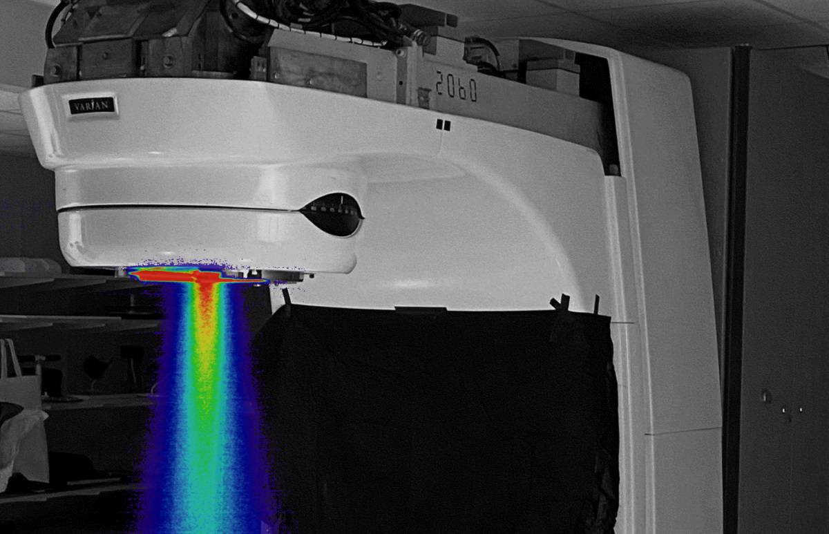
The researchers converted a Varian Clinac 2100 C/D to deliver ultrahigh-dose rate electron beams. The converted system could achieve dose rates of up to 290±5 Gy/s at the isocentre (at a source-to-surface distance of 100 cm), well above the reported 40 Gy/s threshold needed to potentially achieve the FLASH effect. They note that after conversion, the beam can be used in a conventional geometry with the patient on the treatment couch. The group is using the ultrahigh-dose rate beam in preclinical studies, as well as in clinical veterinary treatments of dogs with sarcoma tumours.
A disappearing pacemaker
Temporary cardiac pacemakers provide pacing for patients with short-term heart rhythm disorders. But such devices typically use leads inserted through the skin and require surgical removal when no longer needed. A team headed up at Northwestern University and The George Washington University has developed an implantable cardiac pacemaker with no external leads that provides post-operative control of heart rate and rhythm, and then completely dissolves in the body after completion of therapy.
The pacemaker is made entirely from bioresorbable materials, and receives power and control commands via wireless energy transfer. The researchers demonstrated successful pacing in ex vivo animal hearts and human cardiac tissue, and in vivo in a dog during open-chest surgery. They also implanted the pacemakers in rats and performed daily pacing trials on the animals, observing successful ventricular capture.
First positronium image recorded during a PET scan
Positron emission tomography (PET) is a molecular imaging tool commonly employed for cancer diagnosis. During a PET scan, positronium is also generated in the patient’s body, but current PET systems are not able to acquire positronium images. A research group headed up at Jagiellonian University has now created the first-ever positronium image recorded during a PET scan. Their goal is to use positronium imaging to distinguish between healthy, cancerous and inflammatory tissues.
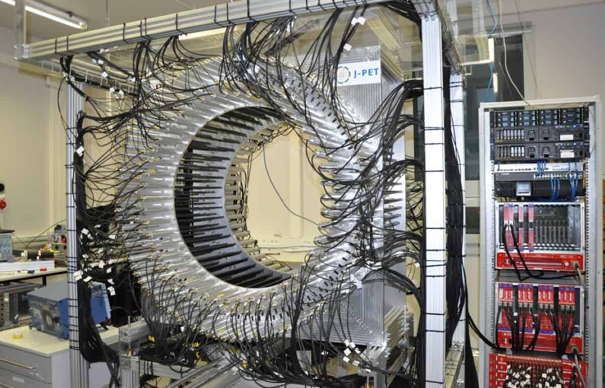
The researchers developed a method for positronium lifetime imaging using the Jagiellonian-PET (J-PET) scanner, which is based on low-cost plastic scintillators arranged in concentric layers and read out by photomultipliers. Images of cancerous and healthy tissue samples exhibited significant differences in positronium lifetime, suggesting that this approach could help determine the degree of cancer malignancy in vivo without the need for surgical biopsy. Next, the team aims to construct a high-sensitivity total-body J-PET and perform positronium imaging in vivo.
The world’s first photon-counting CT scanner
Computed tomography, or CT, is a ubiquitous X-ray imaging technique used to perform more than 300 million medical imaging exams globally each year. Now, following than 15 years of research, Siemens Healthineers has unveiled the world’s first photon-counting CT scanner, the Naeotom Alpha.
Conventional CT requires two conversion steps to create a medical image: X-ray photons collected by a scintillator are used to generate an optical signal, which is then converted into an electrical signal. Photon counting CT, however, enables one-step conversion straight from X-ray photons into an electrical current. This provides dramatic imaging improvements, including increased resolution and a reduction in radiation dose by up to 45% over conventional CT detectors. Photon counting technology also makes it possible to assess the energy level of each and any photon separately.

Physics World‘s Breakthrough of the Year coverage is supported by Bluefors, a leading supplier of cryogen-free dilution refrigerator measurement systems with a strong focus on the quantum computing and information community. Our aim is to deliver the most reliable and easy to operate systems on the market which are of the highest possible quality.