Metal implants in the body, such as hip replacements or dental fillings, are a major source of artefacts in CT images. Clinically available methods reduce these metal artefacts – up to a point.
Iterative metal artefact reduction (iMAR) decreases the influence of metal during image reconstruction by interpolating between boundaries of metal in a tissue-normalized sinogram. However, iMAR doesn’t incorporate spectral information, so some anatomic information is lost. A second technique, virtual monoenergetic imaging (VMI), combines multiple spectra from a dual-energy CT acquisition.
New photon-counting CT systems inherently provide spectral information without requiring a dual-energy acquisition and offer better spatial resolution, improved contrast-to-noise ratio and lower radiation dose than conventional energy-integrating detectors. The first photon-counting CT system, the NAEOTOM Alpha from Siemens Healthineers, received CE certification and 510(k) FDA clearance in 2021.
Julian Anhaus, a third-year PhD student in the CT Physics department at Siemens Healthineers in Germany, is investigating different approaches for metal artefact reduction on these systems. He and his PhD research advisors, Christian Hofmann, a global CT technology manager at Siemens Healthineers, and Andreas Mahnken, a professor at Philipps-Universität Marburg, recently studied some of the possibilities for metal artefact reduction on the NAEOTOM Alpha, publishing their findings in Physics in Medicine & Biology.
“Of course, clinicians must make their own experiences with different protocols and consequentially spectra and other scan parameters, patients and implant types, but this work can be used as an orientation and guideline for clinicians on how to tune their clinical protocols on the NAEOTOM Alpha when performing scans on patients with metal to provide best possible images for the patient’s diagnosis or care,” Anhaus says.
VMI plus iMAR reduces artefacts from high-Z metals
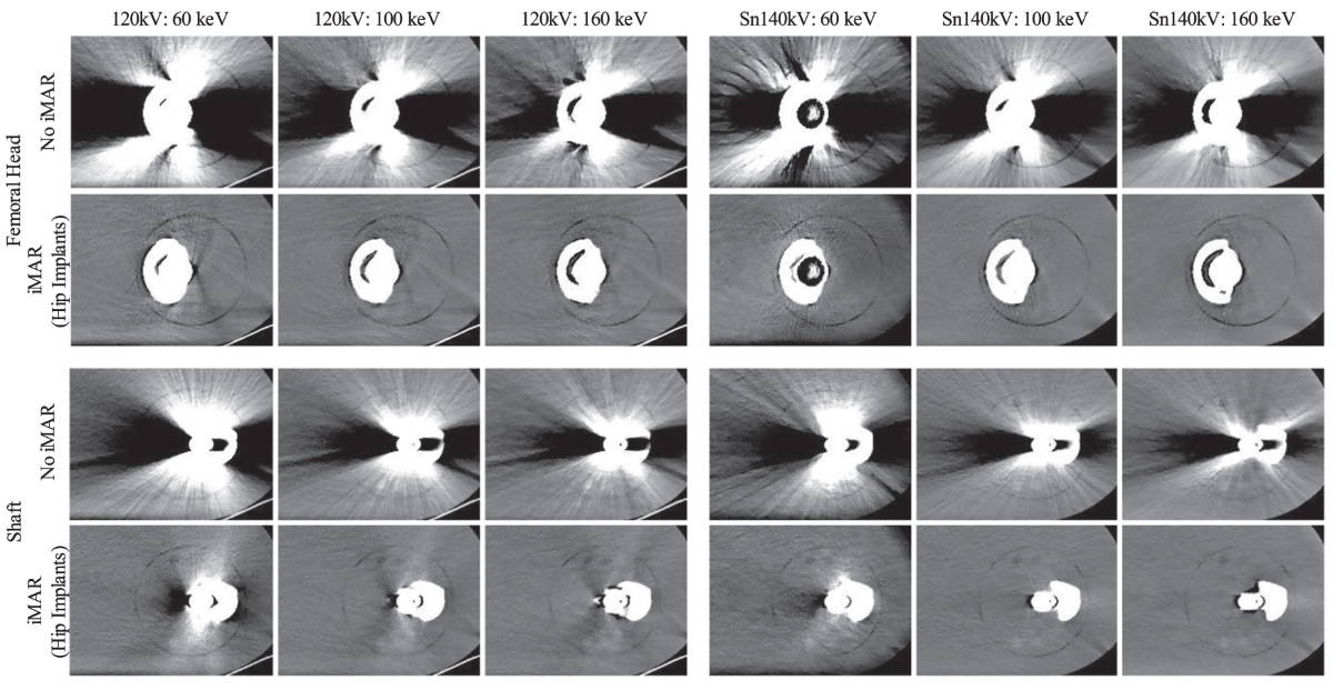
Anhaus tested iMAR and VMI on the NAEOTOM Alpha using anthropomorphic phantoms for different body regions and a tissue characterization phantom. He reconstructed images with and without iMAR and computed VMIs in 10 keV steps from 40 to 190 keV.
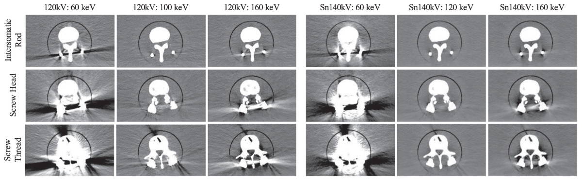
Results were mixed – Anhaus found that VMI could reduce metal artefacts in metals with low atomic numbers and low penetration lengths, such as those used in spinal implants. But for cases with large metal implants and materials with high atomic numbers, such as those used in dental fillings or in the hip head, he could only reduce artefacts by applying iMAR after VMI.
“The inherent spectral information, which is one of the main benefits of recent [photon-counting detector CT] systems, can be utilized to reduce metal artefacts without iMAR. Unfortunately, this only applies to certain metal and implant types,” Anhaus summarizes. “In most implant types, the utilization of spectral information reduces the base artefact level, but iMAR is still required to provide metal artefact-free CT images.”
Anhaus says that this study only scratches the surface of metal artefact reduction options for the NAEOTOM Alpha and other photon-counting CT systems.
Deep learning transforms standard CT scans towards spectral images
“There are many thrilling methods which can be enabled through the photon-counting detector system and carried over to [metal artefact reduction] applications,” he says.
He and Hofmann emphasize that this study is not intended to compare image quality and artefact reduction potential between photon-counting CT and conventional energy-integrating detector CT systems that require dual-energy CT acquisition for VMI. A comprehensive comparison is part of their future work.