Increasingly complex research questions demand increasingly complex data – of which 3D data is one example. The move from 2D to 3D gives us (quite literally) another dimension to work with. PhD candidate Sara Ryding explains how ecological 3D data can reveal previously hidden knowledge.
3D data adds an extra layer to traditional 2D measurements by capturing oddities in curves and depth which may otherwise not be appreciated. The most widely used application of 3D data is shape analysis, usually in the form of geometric morphometrics (i.e., how does “maths” describe this shape). However, this is not its only use, as 3D data can also store valuable information on size, surface texture and colour.
The use of 3D data in ecology is steadily increasing but remains in its infancy because 3D methods are still relatively new and largely inaccessible to many researchers. Therefore, a lot of the current focus has been placed on verifying methods and identifying differences between species.
The technology
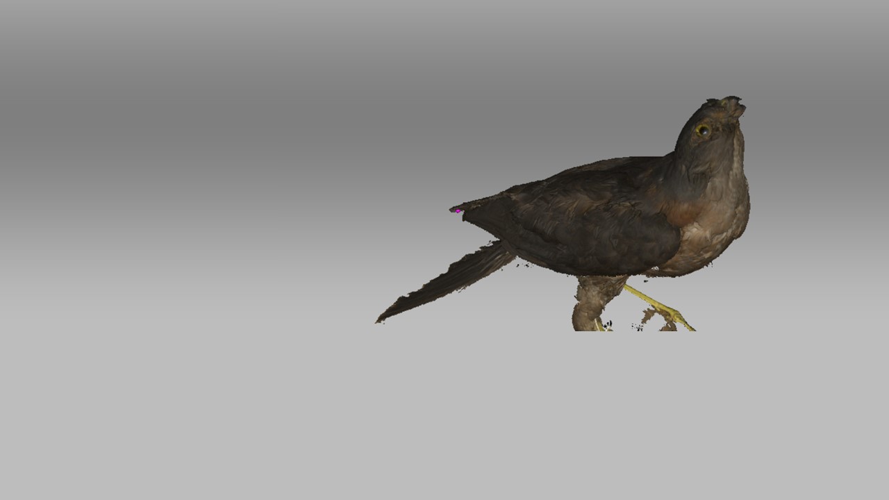
At the forefront of 3D data capture are CT and 3D scanning. While 3D scanning digitises the external surface of the structure, CT scanning can go beyond this to capture internal structures.
However, there’s a trade-off – CT scanning requires a stationary (and often large) machine, whereas 3D scanning can be done with a portable device. This difference in mobility also influences how efficient the methods are; a 3D surface scan can be done in 10-15 minutes, whereas CT scans can take from 20 minutes up to an hour.
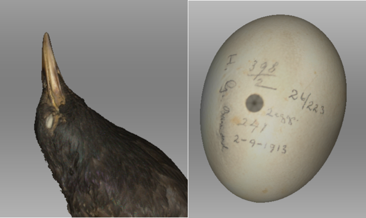
Both methods show great promise in improving 2D-based estimates. For example, the use of micro CT scanning has supported traditional 2D estimates of size of bird bills, whereas others have used 3D data to show that 2D methods may underestimate variability in egg shape.
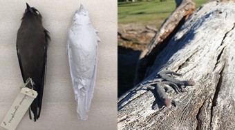
Because the object being digitised needs to be stationary during image acquisition, 3D digitization is often done on a deceased specimen. Of course, if you’re an ecologist looking for a bunch of dead animals, there’s a very good place to start – the museum.
Museums and 3D digitization
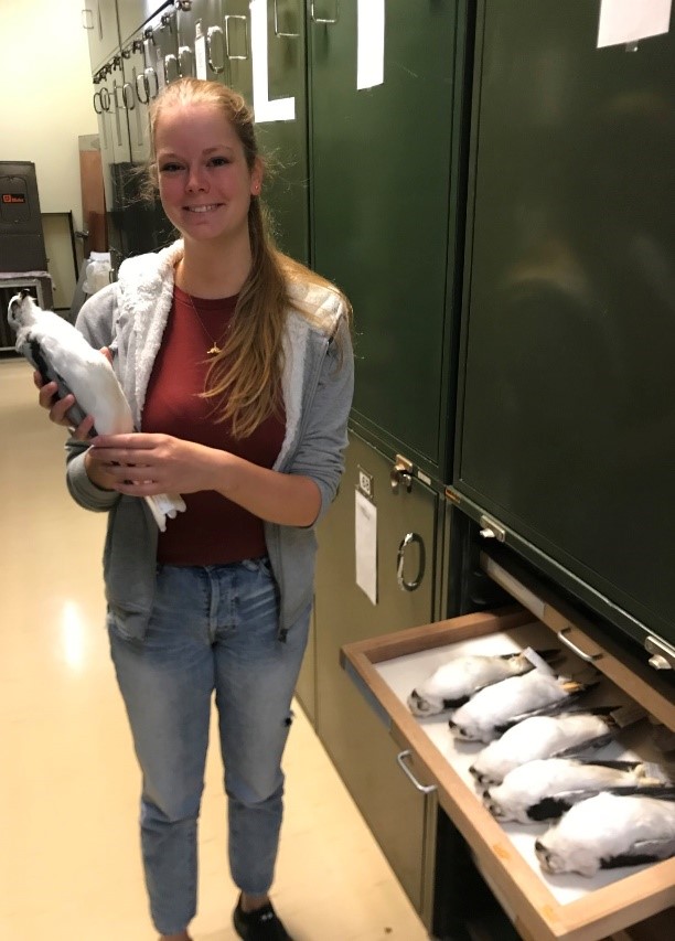
A collection vault full of thousands of dead animals may not be the first thing that comes to mind when you think “applied ecology”. However, these repositories hold valuable information on species distributions, phenotypes, genotypes and environmental pollution, to name a few.
The importance of museum collections has been stressed intermittently for the past few decades amidst dwindling financial support. The new frontier of this is an emphasis on digitising museums, seen in part by new funding initiatives that focus specifically on digitising collections.
This is where 3D digitisation can really shine: the methods are non-invasive and non-damaging, the data can be used in a myriad of different ways, and the 3D files produced can be used freely by researchers, thereby protecting valuable specimens from future (potentially harmful) manual handling.
3D data for Ecology
Work is ongoing to determine how best to use 3D data to answer ecological questions. Most current studies are focusing on shape and associations between morphology and ecology, both in extant and recently extinct animals. There is also a focus on the evolution of traits and ecological patterns which occasionally incorporate citizen science.
My own research is concerned with size and surface area, rather than shape. I plan on using 3D data to investigate changes in size within and between species, relating this to their thermal environment (i.e., climate change).
In addition to using 3D data for size and shape analysis, 3D printing holds many potential ecological applications. High fidelity 3D prints can be used to study otherwise difficult ecological interactions in more controlled settings, such as that between flowers and bees. Prints can also be used to study predator attack rates against their prey, similar to clay models (but 3D prints can be produced in faster and with more morphological accuracy), and for physiological studies on thermal ecology.
I am so excited to be using 3D scanners and data in my work and can’t wait to see how 3D data will be used in the future to improve ecological research.
Have any experience working with 3D scanners or would like to incorporate it in your work? Share your thoughts in the comments below.
Bio
Sara Ryding, Deakin University
Sara is a PhD Candidate studying changes in bill and leg size of birds in response to climate change over the past 100 years, looking at a wide range of species and a wide distribution across Australia. The project will try to answer several important questions, including to what extent birds are changing morphology in response to climatic warming, what factors are causing these changes, and why some birds are or are not changing.
Follow Sara @zuuletc
