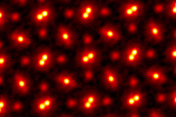
Behold the highest-resolution image of atoms ever taken. To create it, Cornell University researchers captured a sample from a crystal in three dimensions and magnified it 100 million times, doubling the resolution that earned the same scientists a Guinness World Record in 2018. Their imaging process could help develop materials for designing more powerful and efficient phones, computers and other electronics, as well as longer-lasting batteries.
The scientists obtained the image using a technique called electron ptychography. It involves shooting a beam of electrons, about a billion per second, at a target material. The beam moves infinitesimally as the electrons are fired, so they hit the sample from slightly different angles—sometimes they pass through cleanly; other times they collide with atoms and bounce around inside the sample before exiting. Cornell physicist David Muller likens the technique to playing dodgeball against opponents who are standing in the dark. The dodgeballs are electrons, and their targets are individual atoms. Although Muller cannot see the targets, he can detect where the “dodgeballs” end up. Based on the speckle pattern generated by billions of these electrons as they hit a detector, machine-learning algorithms can calculate where the atoms were in the sample and what their shapes might be, thus creating an image.
Previously, electron ptychography had only been used to image extremely flat samples just one to a few atoms thick. But Muller and his colleagues' new study in Science describes capturing multiple layers tens to hundreds of atoms thick. This makes the technique much more relevant to materials scientists, who typically study the properties of samples with a thickness of about 30 to 50 nanometers. (This is smaller than the length your fingernails grow in a minute but many times thicker than what electron ptychography could image in the past.) “They can actually look at stacks of atoms now, so it's amazing,” says University of Sheffield engineer Andrew Maiden, who helped to develop ptychography but was not part of the new study. “The resolution is just staggering.”
On supporting science journalism
If you're enjoying this article, consider supporting our award-winning journalism by subscribing. By purchasing a subscription you are helping to ensure the future of impactful stories about the discoveries and ideas shaping our world today.
This result marks an important advancement in the world of electron microscopy. Invented in the early 1930s, standard electron microscopes made it possible to see objects such as polioviruses, which are smaller than the wavelengths of visible light. But electron microscopes had a limit: increasing their resolution required raising the electron beam's energy, and eventually the necessary energy would become so great that it would damage the sample.
Ptychography, in contrast, uses a detector that can record all the different angles the beam can scatter to at every beam position, getting much more information with the same wavelength and lens. Researchers theorized ptychography in the 1960s and conceived its use to overcome electron lenses' limits in the 1980s. But because of computing and detector limitations and the complex math required, the technique was not put into practice for decades. Early versions worked far better with visible light and x-rays than the electrons needed to image atomic-size objects. Meanwhile scientists kept improving electron microscopes. “You had to be a true believer in ptychography to be paying attention to it,” Muller says.
Just in the past several years Muller and his team developed a detector good enough for electron ptychography to work experimentally. By 2018 they had figured out how to reconstruct two-dimensional samples with the technique, producing what Muller calls “the highest-resolution image by any method in the world” (and winning that Guinness record). The researchers accomplished this feat using a lower-energy wavelength than other methods, letting them better preserve what they viewed.
The next challenge was thicker samples, in which an electron wave ricochets off many atoms before reaching a detector: the so-called multiple scattering problem. The team members found that with enough overlapping speckle patterns and computing power (and, according to Muller, “brute force and ignorance”), they could work backward to determine which layout of atoms produced a given pattern. To do this, they fine-tuned a model until the pattern it generated matched the experimentally produced one.
Such high-resolution imaging techniques are essential for developing the next generation of electronic devices. For example, many researchers are looking beyond silicon-based computer chips to find more efficient semiconductors. To make this happen, engineers need to know what they are working with at an atomic level—which means using technologies such as electron ptychography. “We have these tools sitting there, waiting to help us optimize what will become the next generation of devices,” says J. Murray Gibson, dean of the Florida A&M University–Florida State University College of Engineering, who was not part of the new study.
Batteries are a particularly promising area for applying imaging techniques such as electron ptychography, says Roger Falcone, a physicist at the University of California, Berkeley, who was also not involved with the research. Making batteries that can store a lot of energy safely is critical for the transition from fossil fuels to renewable energies, including wind and solar. “Imaging technologies are very important to improving batteries because we can look at the chemical reactions in detail,” Falcone says.
But there is still a long way to go. For electron ptychography to lead to breakthroughs for your cell phone or laptop, it must do more than reconstruct an image—it must precisely locate an individual atom in a material. Although the scientists showed how their new process could do so in theory, they have not yet demonstrated it experimentally. “With any new technique, it always takes a bit of time for your fellow researchers to try this out and see if it bears out into real, practical uses,” says Leslie Thompson, a materials characterization expert at IBM, who was not involved in the new study.
“To the extent that you invent a new tool like a high-resolution microscope, my sense is you tend to be surprised [by] what problem it's applied to solve,” Falcone says. “People will look at things we can't even imagine now—and solve a problem that we're not even sure we have yet.”